

搜索
请登录


提醒成功


 微信/QQ登录
搜索


 首页
首页
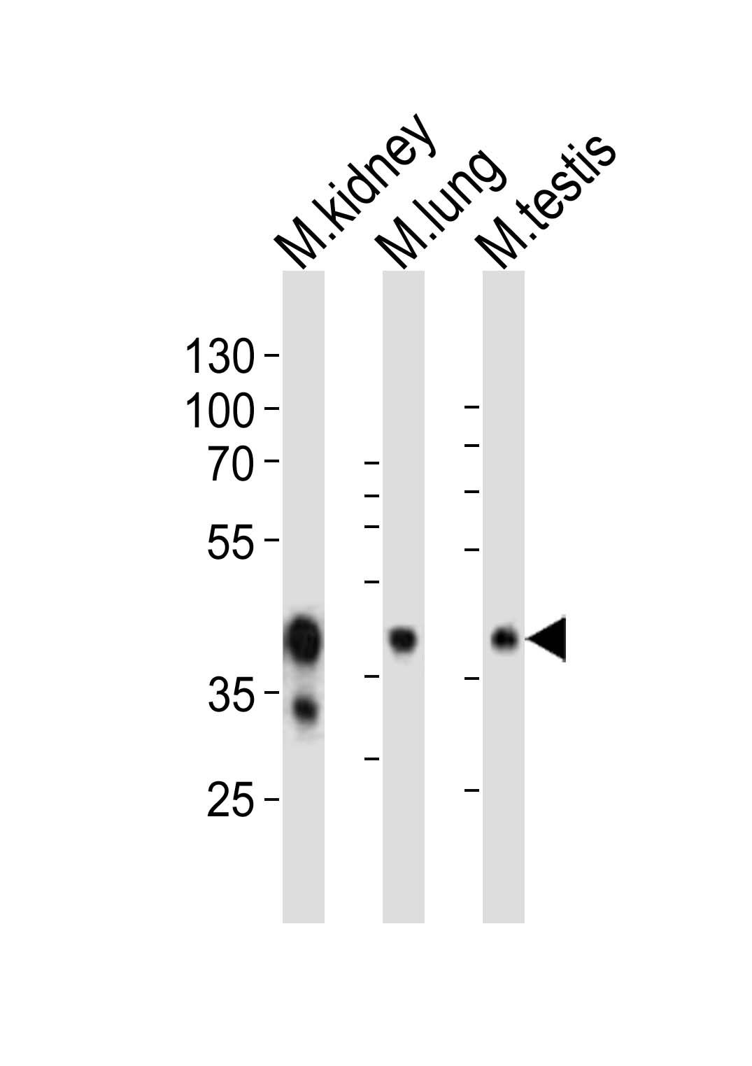
Western blot analysis of lysates from mouse kidney, mouse lung, mouse testis tissue lysate (from left to right), using Epcam Antibody (C-term)(Cat. #P32953). P32953 was diluted at 1:1000 at each lane. A goat anti-rabbit IgG H&L(HRP) at 1:10000 dilution was used as the secondary antibody. Lysates at 20ug per lane.
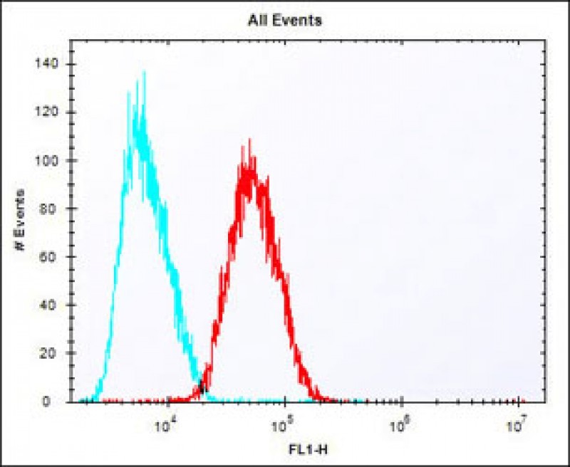
Overlay histogram showing HepG2 cells stained with P32953 (red line). The cells were fixed with 2% paraformaldehyde (10 min) and then permeabilized with 90% methanol for 10 min. The cells were then icubated in 2% bovine serum albumin to block non-specific protein-protein interactions followed by the antibody (P32953, 1:25 dilution) for 60 min at 37ºC. The secondary antibody used was Alexa Fluor® 488 goat anti-rabbit lgG (H+L) (1583138) at 1/400 dilution for 40 min at 37ºC. Isotype control antibody (blue line) was rabbit IgG1 (1μg/1x10^6 cells) used under the same conditions. Acquisition of >10, 000 events was performed.
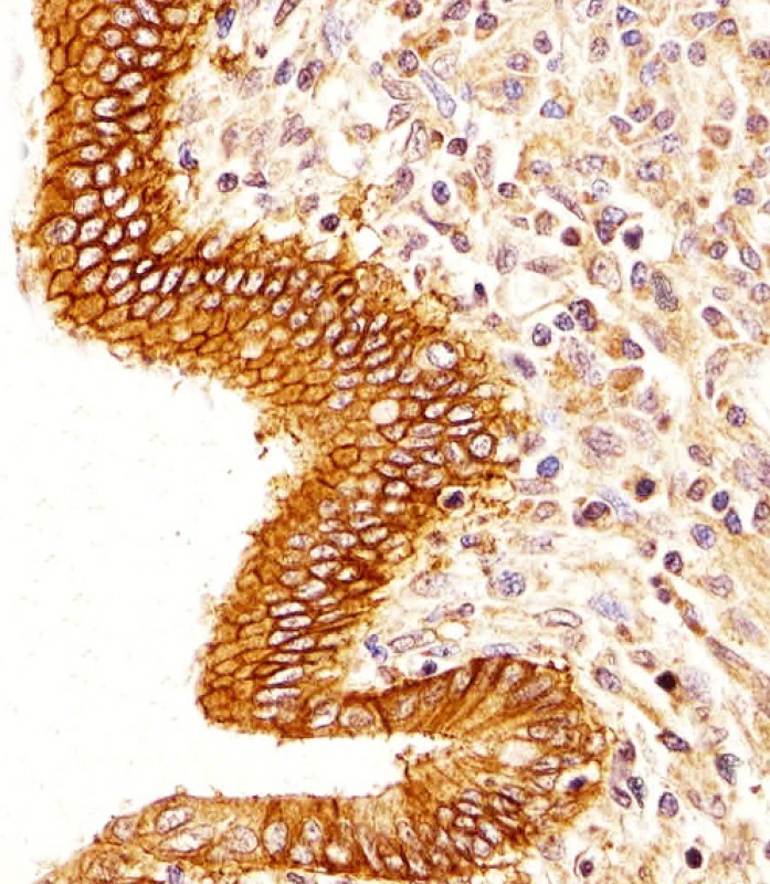
P32953 staining Epcam in Human colorectal carcinoma tissue sections by Immunohistochemistry (IHC-P - paraformaldehyde-fixed, paraffin-embedded sections). Tissue was fixed with formaldehyde and blocked with 3% BSA for 0. 5 hour at room temperature; antigen retrieval was by heat mediation with a citrate buffer (pH6). Samples were incubated with primary antibody (1/25) for 1 hours at 37°C. A undiluted biotinylated goat polyvalent antibody was used as the secondary antibody.
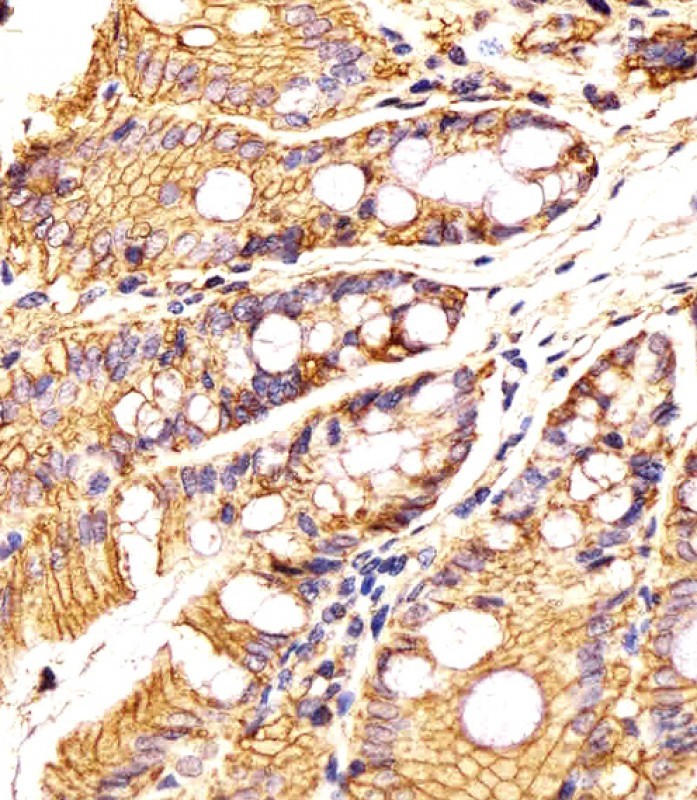
P32953 staining Epcam in Mouse colon tissue sections by Immunohistochemistry (IHC-P - paraformaldehyde-fixed, paraffin-embedded sections). Tissue was fixed with formaldehyde and blocked with 3% BSA for 0. 5 hour at room temperature; antigen retrieval was by heat mediation with a citrate buffer (pH6). Samples were incubated with primary antibody (1/25) for 1 hours at 37°C. A undiluted biotinylated goat polyvalent antibody was used as the secondary antibody.
Rabbit Polyclonal Antibody to (Mouse) Epcam
-
货号:
P32953 -
别名:
Epithelial cell adhesion molecule, Ep-CAM, Epithelial glycoprotein 314, EGP314, mEGP314, Protein 289A, Tumor-associated calcium signal transducer 1, CD326, Epcam, Tacstd1 -
应用:
WB,IHC,FCM -
反应种属:
Human, Mouse, Rat -
抗体类型:
Primary antibody -
Swissprot:
Q99JW5 -
规格:
-
数量:
-+ -
说明书:

目录价¥1980


Rabbit Polyclonal Antibody to (Mouse) Epcam
Description |
|---|
May act as a physical homophilic interaction molecule between intestinal epithelial cells (IECs) and intraepithelial lymphocytes (IELs) at the mucosal epithelium for providing immunological barrier as a first line of defense against mucosal infection. Plays a role in embryonic stem cells proliferation and differentiation. Up-regulates the expression of FABP5, MYC and cyclins A and E (By similarity). |
Specification |
|
|---|---|
| Aliases | Epithelial cell adhesion molecule, Ep-CAM, Epithelial glycoprotein 314, EGP314, mEGP314, Protein 289A, Tumor-associated calcium signal transducer 1, CD326, Epcam, Tacstd1 |
| Entrez GeneID | 17075 |
| Swissprot | Q99JW5 |
| WB Predicted band size | 35.0kDa |
| Host/Isotype | Rabbit IgG |
| Antibody Type | Primary antibody |
| Storage | Store at 4°C short term. Aliquot and store at -20°C long term. Avoid freeze/thaw cycles. |
| Species Reactivity | Human, Mouse, Rat |
| Immunogen | This mouse Epcam antibody is generated from a rabbit immunized with a KLH conjugated synthetic peptide between 302-335 amino acids from the C-terminal region of mouse Epcam. |
Application |
|
|---|---|
| WB | 1/1000 |
| IHC | 1/100-1/500 |
| FCM | 1/25 |
Product Image
- Western blot analysis of lysates from mouse kidney, mouse lung, mouse testis tissue lysate (from left to right), using Epcam Antibody (C-term)(Cat. #P32953). P32953 was diluted at 1:1000 at each lane. A goat anti-rabbit IgG H&L(HRP) at 1:10000 dilution was used as the secondary antibody. Lysates at 20ug per lane.
- Overlay histogram showing HepG2 cells stained with P32953 (red line). The cells were fixed with 2% paraformaldehyde (10 min) and then permeabilized with 90% methanol for 10 min. The cells were then icubated in 2% bovine serum albumin to block non-specific protein-protein interactions followed by the antibody (P32953, 1:25 dilution) for 60 min at 37ºC. The secondary antibody used was Alexa Fluor® 488 goat anti-rabbit lgG (H+L) (1583138) at 1/400 dilution for 40 min at 37ºC. Isotype control antibody (blue line) was rabbit IgG1 (1μg/1x10^6 cells) used under the same conditions. Acquisition of >10, 000 events was performed.
- P32953 staining Epcam in Human colorectal carcinoma tissue sections by Immunohistochemistry (IHC-P - paraformaldehyde-fixed, paraffin-embedded sections). Tissue was fixed with formaldehyde and blocked with 3% BSA for 0. 5 hour at room temperature; antigen retrieval was by heat mediation with a citrate buffer (pH6). Samples were incubated with primary antibody (1/25) for 1 hours at 37°C. A undiluted biotinylated goat polyvalent antibody was used as the secondary antibody.
- P32953 staining Epcam in Mouse colon tissue sections by Immunohistochemistry (IHC-P - paraformaldehyde-fixed, paraffin-embedded sections). Tissue was fixed with formaldehyde and blocked with 3% BSA for 0. 5 hour at room temperature; antigen retrieval was by heat mediation with a citrate buffer (pH6). Samples were incubated with primary antibody (1/25) for 1 hours at 37°C. A undiluted biotinylated goat polyvalent antibody was used as the secondary antibody.
For Reseach Only
Application Key:WB - Western Blot | IHC - Immunohistochemistry | ICC - Immunocytochemistry | FCM - Flow Cytometry | ELISA - Enzyme-linked Immunosorbent Assay | IP - Immunoprecipitation
#P32953

相关产品


联系方式
CONTACT-
联系电话:
0731-88388787 -
公司邮箱:
sales@promab.cn -
公司地址:
湖南省长沙市高新开发区林语路239号顺畅产业园5楼
湖南省长沙市望城区普瑞大道8号出版科技园2号楼
 官方微信
官方微信
产品中心
Product技术服务
service客户留言
message