

搜索
请登录


提醒成功


 微信/QQ登录
搜索


 首页
首页
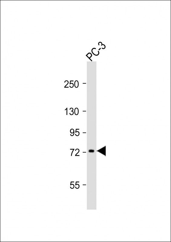
Anti-DVL1 Antibody (Center)at 1:1000 dilution + PC-3 whole cell lysates
Lysates/proteins at 20 µg per lane.
Secondary
Goat Anti-Rabbit IgG, (H+L), Peroxidase conjugated at 1/10000 dilution.
Predicted band size : 75 kDa
Blocking/Dilution buffer: 5% NFDM/TBST.
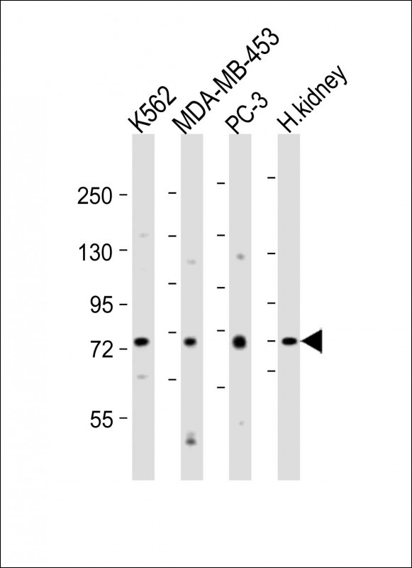
All lanes : Anti-DVL1 Antibody (Center) at 1:2000 dilution
Lane 1: K562 whole cell lysates
Lane 2: MDA-MB-453 whole cell lysates
Lane 3: PC-3 whole cell lysates
Lane 4: human kidney lysates
Lysates/proteins at 20 µg per lane.
Secondary
Goat Anti-Rabbit IgG, (H+L), Peroxidase conjugated at 1/10000 dilution.
Predicted band size : 75 kDa
Blocking/Dilution buffer: 5% NFDM/TBST.
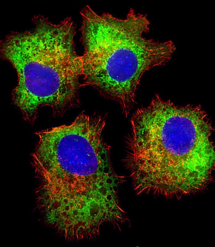
Immunofluorescent analysis of 4% paraformaldehyde-fixed, 0.1% Triton X-100 permeabilized HepG2 (human liver hepatocellular carcinoma cell line) cells labeling DVL1 with P33217 at 1/25 dilution, followed by Dylight® 488-conjugated goat anti-rabbit IgG secondary antibody at 1/200 dilution (green). Immunofluorescence image showing cytoplasm staining on HepG2 cell line. Cytoplasmic actin is detected with Dylight® 554 Phalloidin at 1/100 dilution (red).The nuclear counter stain is DAPI (blue).
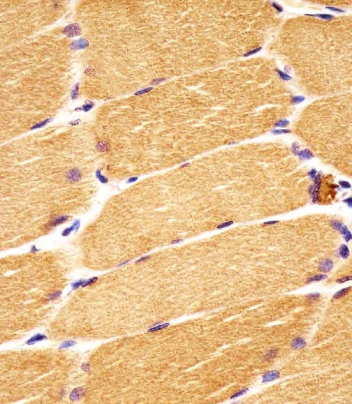
P33217 staining DVL1 in human skeletal muscle sections by Immunohistochemistry (IHC-P - paraformaldehyde-fixed, paraffin-embedded sections). Tissue was fixed with formaldehyde and blocked with 3% BSA for 0. 5 hour at room temperature; antigen retrieval was by heat mediation with a citrate buffer (pH6). Samples were incubated with primary antibody (1/25) for 1 hours at 37°C. A undiluted biotinylated goat polyvalent antibody was used as the secondary antibody.
Rabbit Polyclonal Antibody to DVL1
-
货号:
P33217 -
别名:
Segment polarity protein dishevelled homolog DVL-1, Dishevelled-1, DSH homolog 1, DVL1 -
应用:
WB,IHC,IF -
反应种属:
Human, Mouse, Rat -
抗体类型:
Primary antibody -
Swissprot:
O14640 -
规格:
-
数量:
-+ -
说明书:

目录价¥1980


Rabbit Polyclonal Antibody to DVL1
Description |
|---|
DVL1, the human homolog of the Drosophila dishevelled gene (dsh) encodes a cytoplasmic phosphoprotein that regulates cell proliferation, acting as a transducer molecule for developmental processes, including segmentation and neuroblast specification. DVL1 is a candidate gene for neuroblastomatous transformation. The Schwartz-Jampel syndrome and Charcot-Marie-Tooth disease type 2A have been mapped to the same region as DVL1. The phenotypes of these diseases may be consistent with defects which might be expected from aberrant expression of a DVL gene during development. |
Specification |
|
|---|---|
| Aliases | Segment polarity protein dishevelled homolog DVL-1, Dishevelled-1, DSH homolog 1, DVL1 |
| Entrez GeneID | 1855 |
| Swissprot | O14640 |
| WB Predicted band size | 75.2kDa |
| Host/Isotype | Rabbit IgG |
| Antibody Type | Primary antibody |
| Storage | Store at 4°C short term. Aliquot and store at -20°C long term. Avoid freeze/thaw cycles. |
| Species Reactivity | Human, Mouse, Rat |
| Immunogen | This DVL1 antibody is generated from rabbits immunized with a KLH conjugated synthetic peptide between 442-470 amino acids from the Central region of human DVL1. |
| Formulation | Purified antibody in PBS with 0.05% sodium azide. |
Application |
|
|---|---|
| WB | 1/1000-1/2000 |
| IHC | 1/100-1/500 |
| IF | 1/25 |
Product Image
- Anti-DVL1 Antibody (Center)at 1:1000 dilution + PC-3 whole cell lysates Lysates/proteins at 20 µg per lane. Secondary Goat Anti-Rabbit IgG, (H+L), Peroxidase conjugated at 1/10000 dilution. Predicted band size : 75 kDa Blocking/Dilution buffer: 5% NFDM/TBST.
- All lanes : Anti-DVL1 Antibody (Center) at 1:2000 dilution Lane 1: K562 whole cell lysates Lane 2: MDA-MB-453 whole cell lysates Lane 3: PC-3 whole cell lysates Lane 4: human kidney lysates Lysates/proteins at 20 µg per lane. Secondary Goat Anti-Rabbit IgG, (H+L), Peroxidase conjugated at 1/10000 dilution. Predicted band size : 75 kDa Blocking/Dilution buffer: 5% NFDM/TBST.
- Immunofluorescent analysis of 4% paraformaldehyde-fixed, 0.1% Triton X-100 permeabilized HepG2 (human liver hepatocellular carcinoma cell line) cells labeling DVL1 with P33217 at 1/25 dilution, followed by Dylight® 488-conjugated goat anti-rabbit IgG secondary antibody at 1/200 dilution (green). Immunofluorescence image showing cytoplasm staining on HepG2 cell line. Cytoplasmic actin is detected with Dylight® 554 Phalloidin at 1/100 dilution (red).The nuclear counter stain is DAPI (blue).
- P33217 staining DVL1 in human skeletal muscle sections by Immunohistochemistry (IHC-P - paraformaldehyde-fixed, paraffin-embedded sections). Tissue was fixed with formaldehyde and blocked with 3% BSA for 0. 5 hour at room temperature; antigen retrieval was by heat mediation with a citrate buffer (pH6). Samples were incubated with primary antibody (1/25) for 1 hours at 37°C. A undiluted biotinylated goat polyvalent antibody was used as the secondary antibody.
For Reseach Only
Application Key:WB - Western Blot | IHC - Immunohistochemistry | ICC - Immunocytochemistry | FCM - Flow Cytometry | ELISA - Enzyme-linked Immunosorbent Assay | IP - Immunoprecipitation
#P33217

相关产品


联系方式
CONTACT-
联系电话:
0731-88388787 -
公司邮箱:
sales@promab.cn -
公司地址:
湖南省长沙市高新开发区林语路239号顺畅产业园5楼
湖南省长沙市望城区普瑞大道8号出版科技园2号楼
 官方微信
官方微信
产品中心
Product技术服务
service客户留言
message